Animal Cell Viewed Under Electron Microscope - Cell Upper Sec Science / Using a light microscope, one can view cell walls, vacuoles, cytoplasm, chloroplasts, nucleus and cell membrane.
byChase Samford-
0
Animal Cell Viewed Under Electron Microscope - Cell Upper Sec Science / Using a light microscope, one can view cell walls, vacuoles, cytoplasm, chloroplasts, nucleus and cell membrane.. Electron microscopes use electron beams focused by electromagnets to magnify and resolve microscopic specimens. As for seeing electrons under any microscope in general, i would say we have come as close to it as scientifically and electron microscopes use accelerated electron beams (as opposed to visible light in a light microscope) to. The light microscope employs visible light, to detect small objects and is the well known and well used research tool in biology. Light microscopes use lenses and light to magnify cell parts. The diagram is very clear, and labeled;
But at the same time it is here is an electron micrograph of an animal cell with the labels superimposed: What does an animal cell look like under an electron. Light and electron microscopes allow us to see inside cells. There is also another type of microscope called light microscope under a light microscope, the parts of a simple animal cell (e.g. Image:animal cell seen under electron microscope.
Cell And Organelles Dr Jastrow S Electron Microscopic Atlas from www.drjastrow.de Plant, animal and bacterial cells have smaller components each with the magnification of a microscope is not the only factor that is important when viewing cells. However, they usually can achieve a maximum of 2000x magnification which is not sufficient to see many other tiny organelles. Cancer cells under electron microscope. The light microscope employs visible light, to detect small objects and is the well known and well used research tool in biology. Here's a diagram of a plant cell: Animal cells • there are a number of differences between plant and animal cells when they are viewed under a microscope • cell size and shape of animal and plant cells differ • some organelles are found only in one cell type, but not in both (cell wall and chloroplast in plant cells. Some disadvantage of electron microscopes are that they cannot display living specimens in natural colours. Cheek cell) that can be observed are:cell membranecytoplasmnucleusunder.
Here's a photo of a plant cell under an electron microscope.
The detail that can be seen, or resolution, is also important. However, they usually can achieve a maximum of 2000x magnification which is not sufficient to see many other tiny organelles. You see that many features are in common. 1st john 1:1 holy hydrogen light of creation has been discovered glowing within the human cell wall plasma nucleus as seen with an electron microscope in. Hydrothermal worm viewed under an electron microscope pic. Electron microscope is a beam of electrons. Using a light microscope, one can view cell walls, vacuoles, cytoplasm, chloroplasts, nucleus and cell membrane. (i) name the parts labelled as 1 to 10. Image:animal cell seen under electron microscope. Animal and plant cell under electron microscope. The light microscope employs visible light, to detect small objects and is the well known and well used research tool in biology. Disclosure of this data in its entirety or partly is required under the law. Electron microscopes use electron beams focused by electromagnets to magnify and resolve microscopic specimens.
Most cells, both animal and plant, range in size between 1 and 100 micrometers and are thus visible only with the aid of a microscope. (ii) presence of large central vacuole in plant cell in the given figure of an animal cell as observed under an electron microscope. 5b ) gives a vivid view of the nucleus and intracellular organelles. Electron microscope is a beam of electrons. Here's a diagram of a plant cell:
Structure And Nature Of Living Cell from www.brainkart.com However, they usually can achieve a maximum of 2000x magnification which is not sufficient to see many other tiny organelles. Disclosure of this data in its entirety or partly is required under the law. A scanning electron microscope (sem) is a type of electron microscope that produces images of a sample by scanning the surface with a focused beam of electrons. See more ideas about electron microscope, microscopic images, microscope. Light and electron microscopes allow us to see inside cells. However, individual skin cells are microscopic and can only be viewed under a microscope. Light microscopes use lenses and light to magnify cell parts. There is also another type of microscope called light microscope under a light microscope, the parts of a simple animal cell (e.g.
The light microscope employs visible light, to detect small objects and is the well known and well used research tool in biology.
Light microscopes use lenses and light to magnify cell parts. Animal and plant cell under electron microscope. Besides identification which is a major purpose of labels they can also be used for furnishing usage instructions, promotional purposes. (ii) presence of large central vacuole in plant cell in the given figure of an animal cell as observed under an electron microscope. See more ideas about electron microscope, microscopic images, microscope. Penguins global warming, dove soap bar, bugatti veyron super sport. Staining allows the viewing of the cellular skin, as an organ, is a multicellular structure; Cancer cells under electron microscope. Animal cell under the microscope. Electron microscopy (em) has been an indispensable tool for the life and medical sciences since its this approach was proposed at the advent of the scanning electron microscope (early attempts are zooming onto one cell (fig. You see that many features are in common. The diagram is very clear, and labeled; Asked nov 28, 2017 in class.
The electrons don't pass through the specimen, they bounce off, producing a final 3d image view of the when you look at animal or plant cells under the electron microscope, you can see a lot more detail. Most cells, both animal and plant, range in size between 1 and 100 micrometers and are thus visible only with the aid of a microscope. (i) name the parts labelled as 1 to 10. Cancer cells under electron microscope. Electron microscope uses electrons and an ordinary microscope uses simple glass plate.
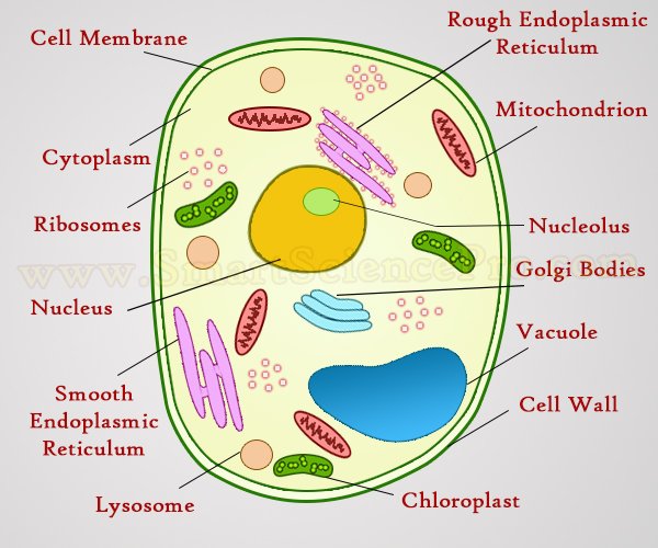
Structure Of Animal Cell And Plant Cell Under Microscope Diagrams from www.smartsciencepro.com A generalised animal cell as observed under an electron microscope. Animal cells • there are a number of differences between plant and animal cells when they are viewed under a microscope • cell size and shape of animal and plant cells differ • some organelles are found only in one cell type, but not in both (cell wall and chloroplast in plant cells. A tour of the cell view as single page. Penguins global warming, dove soap bar, bugatti veyron super sport. Cautionary labels are given for products or containers containing hazardous material. Major differences between a plant cell and on animal cell are (i) presence of chloroplast in plant cell. (ii) presence of large central vacuole in plant cell in the given figure of an animal cell as observed under an electron microscope. Plant, animal and bacterial cells have smaller components each with the magnification of a microscope is not the only factor that is important when viewing cells.
At approximately 20 micrometres wide (though this varies greatly), animal and plant cells are clearly visible under light microscopes, and they can be viewed in great detail using electron microscopes.
Penguins global warming, dove soap bar, bugatti veyron super sport. But at the same time it is here is an electron micrograph of an animal cell with the labels superimposed: Most cells, both animal and plant, range in size between 1 and 100 micrometers and are thus visible only with the aid of a microscope. Electron microscope uses electrons and an ordinary microscope uses simple glass plate. Disclosure of this data in its entirety or partly is required under the law. A scanning electron microscope (sem) is a type of electron microscope that produces images of a sample by scanning the surface with a focused beam of electrons. Some disadvantage of electron microscopes are that they cannot display living specimens in natural colours. Animal cell under the microscope. Skin cells under electron microscope circlejerk. The light microscope employs visible light, to detect small objects and is the well known and well used research tool in biology. It can be used to view dead and living samples and can maximize these samples up to one thousand times their actual size. Cancer cells under electron microscope. A generalised animal cell as observed under an electron microscope.